.svg)
.svg)

.svg)

ترجمه مقاله بارهای ناشی از بافت نرم روی زانوی سالم در فلکشن های فیزیولوژیک مختلف - عددی - نشریه اشپرینگر

عنوان فارسی
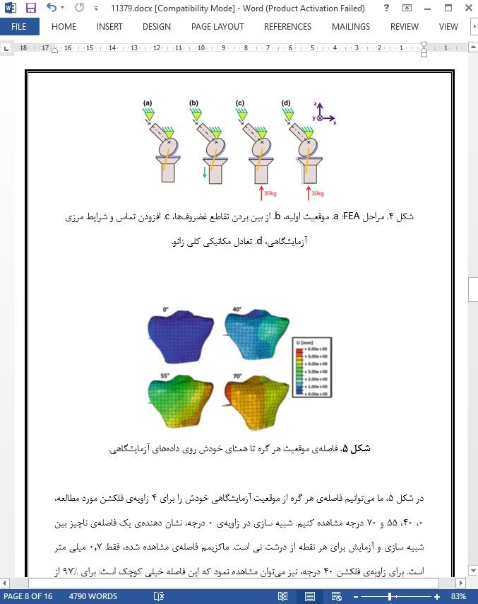
بارهای ناشی از بافت نرم روی زانوی سالم در فلکشن های فیزیولوژیک مختلف: یک رویکرد آزمایشگاهی - عددی
عنوان انگلیسی
Soft Tissues’ Loadings on Healthy Knee at Different Physiological Flexions: A Coupled Experimental–Numerical Approach
صفحات مقاله فارسی
16
صفحات مقاله انگلیسی
14
سال انتشار
2017
رفرنس
دارای رفرنس در داخل متن و انتهای مقاله
نشریه
اشپرینگر - Springer
فرمت مقاله انگلیسی
pdf و ورد تایپ شده با قابلیت ویرایش
فرمت ترجمه مقاله
pdf و ورد تایپ شده با قابلیت ویرایش
فونت ترجمه مقاله
بی نازنین
سایز ترجمه مقاله
14
نوع مقاله
ISI
نوع ارائه مقاله
کنفرانس
شناسه ISSN مجله
2195-271X
کد محصول
11379
وضعیت ترجمه عناوین تصاویر و جداول
ترجمه شده است ✓
وضعیت ترجمه متون داخل تصاویر و جداول
ترجمه نشده است ☓
وضعیت ترجمه منابع داخل متن
ترجمه شده است ✓
ضمیمه
ندارد ☓
بیس
نیست ☓
مدل مفهومی
ندارد ☓
پرسشنامه
ندارد ☓
متغیر
ندارد ☓
رفرنس در ترجمه
در انتهای مقاله درج شده است
رشته های مرتبط با این مقاله
پزشکی و مهندسی پزشکی
گرایش های مرتبط با این مقاله
بیومکانیک، جراحی ارتوپدی
کنفرانس
روش های رایانه ای در بیومکانیک و مهندسی پزشکی - Computer Methods in Biomechanics and Biomedical Engineering
دانشگاه
دانشگاه لیون، فرانسه
doi یا شناسه دیجیتال
https://doi.org/10.1007/978-3-319-59764-5_14
۰.۰
(هنوز امتیازی ثبت نشده است)