ترجمه مقاله نقش ضروری ارتباطات 6G با چشم انداز صنعت 4.0
- مبلغ: ۸۶,۰۰۰ تومان
ترجمه مقاله پایداری توسعه شهری، تعدیل ساختار صنعتی و کارایی کاربری زمین
- مبلغ: ۹۱,۰۰۰ تومان
مقدمه
FMRI بالینی برای برنامهریزی قبل از جراحی
پارادایمهای حسی- حرکتی
پارادایمهای زبانی
پارادایمهای بصری
روایی بالینی و تأثیر جراحی
محدودیتهای BOLD fMRI و نقش تصویربرداری (نقشهبرداری) راکتیویته مغزی عروقی
اهمیت جداشدگی عصبی عروقی
نقشهبرداری CVR
مثال بالینی NVU که با نقشهبرداری BH CVR تشخیص داده شده است
سایر کاربردهای بالینی نقشه برداری CVR
This article provides a review of Blood Oxygen Level Dependent functional magnetic resonance imaging (BOLD fMRI) applications for presurgical mapping in patients with brain tumors who are being considered for lesion resection. Initially, the physical principle of the BOLD effect is discussed, followed by a general overview of the aims of presurgical planning. Subsequently, a review of sensorimotor, language and visual paradigms that are typically utilized in clinical fMRI is provided, followed by a brief description of studies demonstrating the clinical impact of preoperative BOLD fMRI. After this thorough introduction to presurgical fMRI, a detailed explanation of the phenomenon of neurovascular uncoupling (NVU), a major limitation of fMRI, is provided, followed by a discussion of the different approaches taken for BOLD cerebrovascular reactivity (CVR) mapping, which is an effective method of detecting NVU. We then include one clinical case which demonstrates the value of CVR mapping in clinical preoperative fMRI interpretation. The paper then concludes with a brief review of applications of CVR mapping other than for presurgical mapping.
INTRODUCTION
Blood Oxygen Level Dependent functional magnetic resonance imaging (BOLD fMRI) is a brain mapping technique using deoxyhemoglobin contained in the blood vessels as an endogenous contrast agent to produce functional activation maps[1].
Neural activation induces a transient increase in regional oxygen extraction from the blood that is coupled with a much larger increase in cerebral blood flow (CBF) and cerebral blood volume (CBV). This influx of oxygenated hemoglobin results in a net decrease in regional deoxyhemoglobin concentration. This drop in paramagnetic deoxyhemoglobin concentration leads to an increase in the magnetic relaxation times T2 and T2*. The mapping of eloquent areas is thus obtained by acquiring T2 or T2*- weighted images consecutively while the subject is at rest or performing a task, and detecting the signal increase related to the local reduction of deoxyhemoglobin concentration accompanying the functional activation relative to baseline.
OTHER CLINICAL APPLICATIONS OF CVR MAPPING
While CVR is considered a sensitive indicator for assessing the brain’s ability to dynamically adjust its energy supply, the clinical applications of CVR mapping (aside from presurgical mapping) are yet to reach their full potential, mainly due to technical constraints. In recent years, with the development of various advanced approaches, viable clinical applications of CVR mapping have emerged.
CVR mapping in cerebrovascular disease is emerging as a promising tool for clinicians involved in the evaluation of patients at risk for stroke. A few studies have been reported using hypercapnia induced BOLD MRI signal response to probe CVR in patients with arterial steno-occlusive diseases such as carotid artery stenosis[55,71,72]. For example, a patient with right middle cerebral artery occlusion and Moyamoya phenomenon secondary to aplastic anemia showed paradoxical CVR values in the right middle cerebral artery territory in the cortex[55].
این مقاله یک بررسی از کاربردهای تصویربرداری تشدید مغناطیسی عملکردی وابستگی سطح اکسیژن خون (BOLD fMRI) را در نقشهبرداری قبل از جراحی برای برش ضایعه در بیماران مبتلا تومورهای مغزی ارائه میدهد. ابتدا اصل فیزیکی اثر BOLD بحث میشود و سپس یک بررسی کلی از اهداف برنامهریزی قبل از جراحی پرداخته خواهد شد. پس از این الگوهای حسی حرکتی، گفتاری و بینایی بررسی میشود که معمولاً در fMRI بکار گرفته میشود. در ادامه توضیح مختصری از مطالعات بیانگر اثر بالینی BOLD fMRI قبل از جراحی ارائه میشود. بعد از این مقدمه کامل درباره fMRI قبل از جراحی، توضیحی دقیق از پدیده جداشدگی عصبی عروقی (NVU) ارائه میشود که محدودیت اصلی fMRI محسوب میشود و در ادامه رویکردهای متفاوت بکار گرفته شده برای نقشهبرداری راکتیویته مغزی عروقی (CVR) BOLD بحث میشود که روشی مؤثر برای تشخیص NVU هست. آنگاه یک مورد بالینی را آوریم که ارزش نقشهبرداری CVR در تفسیر بالینی fMRI قبل جراحی را نمایان میسازد. در نهایت این مقاله با یک بررسی مختصر از کاربردهای نقشهبرداری CVR در مقایسه با تصویربرداری قبل جراحی نتیجهگیری میکند.
مقدمه
تصویربرداری تشدید مغناطیسی عملکردی وابستگی سطح اکسیژن خون (BOLD fMRI) یک تکنیک تصویربرداری از مغز هست که از دی اکسی هموگلوبین موجود در عروق خونی به عنوان عامل کنتراست دورن زا برای گرفتن نقشههای فعالسازی عملکردی استفاده میشود [1].
فعالسازی نورونی سبب افزایش موقت در تخلیه اکسیژن موضعی از خون میشود که این خون با افزایش خیلی بیشتر در جریان خون مغزی (CBF) و حجم خون مغزی (CBV) وابسته هست. این تخلیه هموگلوبین اکسیژن دار منجر به افت خالص در غلظت دی اکسی هموگلوبین میشود. این افت در غلظت دی اکسی هموگلوبین پارامغناطیس سبب افزایش زمانهای استراحت مغناطیسی T2 و T2* میشود. بنابراین تصویربرداری از مواضع قشر سخنوری از طریق کسب پشت سر هم تصاویر وزندهی شده T2 و T2* بدست آمده است در حالیکه این مسئله در حالت استراحت یا حین انجام وظیفه هست و تشخیص این سیگنال در ارتباط با کاهش موضعی غلظت دی اکسی هموگلوبین همراه با فعالسازی عملکردی نسبت به حالت پایه افزایش مییابد.
سایر کاربردهای بالینی نقشه برداری CVR
در حالیکه CVR به عنوان شاخصی حساس برای ارزشیابی توانایی مغز به تعدیل و تنظیم پویای تأمین انرژی تلقی میشود هنوز کاربردهای بالینی تصویربرداری CVR (بعلاوه نقشهبرداری قبل جراحی) برای تحقق پتانسیل کامل آن عمدتاً به دلیل محدودیتهای فنی حاصل نشده است. در سالیان اخیر، با توسعه رویکردهای پیشرفته مختلف، کاربردهای بالینی قابل اعتماد تصویربرداری CVR ظهور یافتهاند.
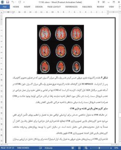
نقشهبرداری CVR در بیماریهای مغزی عروقی به عنوان یک ابزار امیدوارکننده برای پزشکان دخیل در ارزیابی بیماران در معرض خطر سکته مغزی ظهور کرده است. چنین مطالعه با استفاده از هایپرکاپنه ناشی از پاسخ سیگنال BOLD MRI به پروب CVR در بیماران مبتلا به بیماریهای انسداد شریان مانند تنگی شرایین کاروتید گزارش شده است [55،71،72]. به عنوان مثال، بیمار مبتلا به انسداد شریان مغزی راست و پدیده Moyamoya ناشی از آنمی آپلاستیک مقادیر CVR پارادوکسیال را در ناحیه شریان مغز میانی سمت راست در قشر نشان داد [55].