Abstract
Purpose Comorbid depression is common in patients with epilepsy. However, the epilepsy-associated depression is generally atypical and has not been fully recognized by neurologists. This study aimed to compare the behavioral and pathological changes between the chronic lithium chloride-pilocarpine rat epilepsy model (Licl-pilocarpine model) and the Chronic Unpredictable Mild Stress rat depression model (CUMS model), to evaluate for differences between epilepsy-associated depression and primary depression.
Methods The Licl-pilocarpine model and the CUMS model were established respectively and simultaneously. Spontaneous seizures were recorded by video monitoring. Forced swim test (FST) and sucrose consumption test (SCT) were performed to test depressive behaviors. Immobility time (IMT) and climbing time (CMT) in FST, sucrose preference rate (SPR) in SCT, and weight gain rate (WGR) were adopted to represent severity of depressive behaviors in rats. Immunofluorescent staining was conducted to measure expressions of neuronal specific nuclear protein (NeuN), glial fibrillary acidic protein (GFAP), and cluster of differentiation molecule 11b (CD11b) in the hippocampus of Licl-pilocarpine model, CUMS model, and Control group.
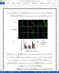
Results Significantly, more prolonged IMT was observed in both the Licl-pilocarpine model (p < 0.05) and the CUMS model (p < 0.01) than Control group. But decreased WGR was only seen in the CUMS model. The percentage of rats with CMT greater than 100 s was significantly higher in the Licl-pilocarpine model than the CUMS model (p < 0.05). Increased CMT was observed in the Licl-pilocarpine model with mild depression subgroup (EMD, IMT ≤ 100 s) even compared with the Control group. Neuronal loss was both found in the Licl-pilocarpine model and the CUMS model when comparing with the Control group (p < 0.05). However, the number of GFAP and CD11b staining cells was both greater in the Licl-pilocarpine model than the CUMS model and the Control group (p < 0.05).
Conclusion There were some different depressive behavioral and hippocampal pathological changes between the Licl-pilocarpine and the CUMS models except for some common features. Gliosis and microglial activation might be more involved in the pathophysiology of epilepsy-associated depression than primary depression.
5. Conclusions
In this study, we used Licl-pilocarpine model and CUMS model to represent epilepsy-associated depression model and primary depression model respectively, and we found that these two models had some different depressive behavioral and hippocampal pathological changes except for some common features. They both had prolonged IMT compared with Control group, but the decreased WGR was only seen in CUMS model, and Licl-pilocarpine model seemed to have more active behaviors even than Control rats. These two models both had hippocampal neuronal loss. However, more prominent gliosis and microglial activation were found in Licl-pilocarpine model than in CUMS model, indicating that gliosis and microglial cell-mediated inflam- matory process might be more greatly involved in epilepsy-associated depression than primary depression.


